Limited-Time Offer: Enjoy 50% Savings! Ends in 00h 00m 00s Coupon code: 50OFF
Skip to content

Free ARDMS Abdomen Sonography Examination AB-Abdomen Exam Questions

Page: 1 / 11 Total 165 questions

Want more questions? Get Premium Access.

Question 1

Which sonographic appearance of the normal epididymis is the most common?

Correct Answer: C. Homogeneous compared to the testis
Explanation:

The normal epididymis typically appears as a homogeneous structure that is either isoechoic or slightly hypoechoic compared to the testis. The most accurate description is 'homogeneous compared to the testis,' meaning the texture is uniform. It is not anechoic, nor does it typically show irregular borders unless pathology is present.

According to Rumack's Diagnostic Ultrasound:

'The normal epididymis appears homogeneous and is isoechoic or slightly hypoechoic relative to the testis.' (Rumack CM et al., Diagnostic Ultrasound, 5th ed.)


Rumack CM, Wilson SR, Charboneau JW, Levine D. Diagnostic Ultrasound. 5th ed. Elsevier; 2017.

AIUM Practice Parameter for Scrotal Ultrasound, 2020.

---
Question 2

What is a major advantage of power Doppler over color flow Doppler?

Correct Answer: C. Improved signal-to-noise ratio
Explanation:

Power Doppler measures the amplitude (strength) of Doppler signals rather than frequency shift, making it more sensitive to low-velocity and small-vessel blood flow. Its primary advantage is an improved signal-to-noise ratio, allowing for better visualization of slow or weak flow.

A: Power Doppler is more sensitive to motion artifacts, not less.

B: It is still angle dependent, though somewhat less so than color Doppler.

D: Power Doppler does not display flow direction (a limitation).

Reference Extracts:

Kremkau FW. Sonography Principles and Instruments. 9th ed. Elsevier, 2015.

Rumack CM, Wilson SR, Charboneau JW, Levine D. Diagnostic Ultrasound. 5th ed. Elsevier, 2017.


Question 3

Which arterial branches lie at the base of the renal pyramids?

Correct Answer: C. Arcuate
Explanation:

The arcuate arteries are located at the corticomedullary junction, arching over the base of the renal pyramids. They form as the interlobar arteries reach the boundary between the cortex and medulla. The arcuate arteries give rise to the interlobular arteries, which supply the renal cortex.

Segmental arteries (A) branch directly from the renal artery.

Interlobar arteries (B) course between the renal pyramids.

Interlobular arteries (D) extend into the cortex from the arcuate arteries.

Reference Extracts:

Moore KL, Dalley AF, Agur AM. Clinically Oriented Anatomy. 7th ed. Lippincott Williams & Wilkins, 2013.

Rumack CM, Wilson SR, Charboneau JW, Levine D. Diagnostic Ultrasound. 5th ed. Elsevier, 2017.

---


Question 4

What is the most common location of a pancreatic pseudocyst?

Correct Answer: A. Lesser sac
Explanation:

Pancreatic pseudocysts most commonly develop in the lesser sac, which lies between the posterior wall of the stomach and the anterior surface of the pancreas. This space allows for the accumulation of pancreatic fluid collections following pancreatitis or pancreatic ductal disruption.

The left anterior pararenal space (B) is a secondary location.

The right subdiaphragmatic space (C) and left pericolic gutter (D) are less common sites.

Reference Extracts:

Mortele KJ, Wiesner W, et al. 'Pancreatic pseudocysts: imaging features and diagnostic difficulties.' Radiographics. 2004;24(4):1005-1020.

Rumack CM, Wilson SR, Charboneau JW, Levine D. Diagnostic Ultrasound. 5th ed. Elsevier, 2017.

---


Question 5

Which pancreatic condition is commonly associated with complete or partial atresia of the duodenum?

Correct Answer: D. Annular pancreas
Explanation:

Annular pancreas is a congenital anomaly in which pancreatic tissue encircles the second part of the duodenum, potentially causing partial or complete duodenal obstruction (atresia). It is due to abnormal migration of the ventral pancreatic bud.

According to Rumack's Diagnostic Ultrasound:

''Annular pancreas results from failure of the ventral pancreatic bud to rotate properly, leading to encirclement of the duodenum.''


Rumack CM, Wilson SR, Charboneau JW, Levine D. Diagnostic Ultrasound. 5th ed. Elsevier, 2017.

Moore KL, Clinically Oriented Anatomy. 8th ed. Wolters Kluwer, 2018.

---

Question 6

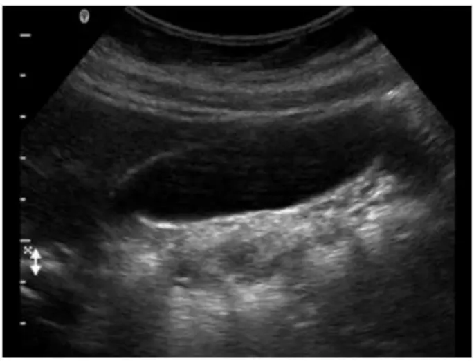
Which artifact is seen within the gallbladder in this image?

Correct Answer: D. Reverberation
Explanation:

The ultrasound image demonstrates multiple parallel echogenic lines within the gallbladder lumen, extending distally and diminishing in intensity. These equidistant lines are classic for a reverberation artifact.

Reverberation occurs when the ultrasound beam reflects multiple times between two strong reflectors (such as the anterior gallbladder wall and the transducer), creating repeating echoes that appear as equally spaced lines. This is frequently seen in:

The gallbladder (especially when collapsed or near the wall)

Near air-filled structures

With metallic objects or surgical clips

Reverberation does not represent a true anatomical structure and should be distinguished from real pathology.

Comparison of answer choices:

A . Banding refers to speed displacement or slice-thickness artifacts and typically does not appear as repetitive lines.

B . Shadowing is a dark band posterior to highly attenuating structures like gallstones or bone, not present here.

C . Attenuation is a general term for the reduction in sound energy as it travels---posterior acoustic shadowing is one type.

D . Reverberation --- Correct. The characteristic bright, parallel lines within the gallbladder lumen confirm this artifact.


Kremkau FW. Sonography Principles and Instruments, 9th ed. Elsevier; 2015.

Rumack CM, Wilson SR, Charboneau JW, Levine D. Diagnostic Ultrasound, 5th ed. Elsevier; 2017.

Hagen-Ansert SL. Textbook of Diagnostic Sonography, 8th ed. Elsevier; 2017.

Question 7

Where in the neck are most thyroid cancer recurrences found?

Correct Answer: B. Ipsilateral
Explanation:

Most thyroid cancer recurrences are found in the ipsilateral neck---particularly in the central (level VI) or lateral (levels II-V) compartments on the same side as the original malignancy.

According to AIUM Practice Parameters:

''Post-thyroidectomy recurrence most frequently occurs ipsilateral to the original tumor, commonly involving regional lymph nodes.''


AIUM Practice Parameter for Thyroid and Neck Ultrasound, 2020.

American Thyroid Association (ATA) Guidelines for Thyroid Cancer Management, 2015.

Question 8

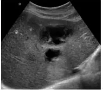
Which abnormality is depicted in this image of a patient who presents with a fever following a liver biopsy?

Correct Answer: C. Abscess
Explanation:

The sonographic image shows a complex fluid collection within the liver parenchyma, with internal echoes and possibly septations, consistent with an abscess. In the clinical context of post-procedural fever following a liver biopsy, a liver abscess is the most likely diagnosis.

A liver abscess appears on ultrasound as a hypoechoic or complex fluid collection that may contain internal debris, septations, or gas (which may produce reverberation artifacts). These features distinguish it from other post-procedural complications.

A cyst (Option A) typically appears as an anechoic, well-defined lesion with posterior acoustic enhancement and no internal debris---this does not match the image or clinical setting.

A biloma (Option B) is a bile collection that can appear similar to a cyst or fluid collection but typically occurs due to bile leak; however, fever and internal complexity on ultrasound more strongly suggest abscess.

A hematoma (Option D) may also appear complex but usually presents with pain and not fever unless secondarily infected. Over time, hematomas evolve in appearance but lack septations and gas unless superinfected.


Rumack, Carol M., et al. Diagnostic Ultrasound. 5th ed., Elsevier, 2018. Chapter: Hepatobiliary System, pp. 107--111.

American Institute of Ultrasound in Medicine (AIUM) Practice Parameter for the Performance of an Ultrasound Examination of the Abdomen and/or Retroperitoneum.

Question 9

When measuring the abdominal aorta, where should the calipers be placed?

Correct Answer: A. Outer wall to outer wall
Explanation:

When measuring the abdominal aorta (or any vessel diameter for aneurysm evaluation), calipers should be placed from outer wall to outer wall to ensure inclusion of the full vessel diameter, including any mural thrombus. This is the standard method accepted by professional societies.

According to AIUM and SRU Guidelines:

''Vessel diameter measurements should be performed from outer wall to outer wall to avoid underestimation of aneurysm size.''


AIUM Practice Parameter for the Performance of Abdominal Aortic Ultrasound, 2020.

Society of Radiologists in Ultrasound (SRU) Consensus Statement, 2003.

---

Question 10

Which of the following must be sterile for a percutaneous procedure?

Correct Answer: D. Gel within transducer cover
Explanation:

In percutaneous procedures such as biopsies or drainages, maintaining a sterile field is critical to avoid introducing infection. While the transducer is covered by a sterile cover, the gel placed inside this cover (between the probe and the cover) must also be sterile, as it contacts the sterile field. The transducer itself and machine controls are not sterile but are handled appropriately to avoid field contamination.

According to the AIUM Guidelines:

'Sterile coupling gel must be used inside the sterile probe cover during all invasive or percutaneous procedures.' (AIUM Guidelines for Cleaning and Preparing Ultrasound Transducers, 2021).


AIUM Guidelines for Cleaning and Preparing Ultrasound Transducers and Equipment for Reuse, 2021.

ACR Practice Parameter for Performing Ultrasound-Guided Procedures, 2020.

---

Question 11

Where is the main pancreatic duct located?

Correct Answer: D. Medial to the superior mesenteric vein
Explanation:

The main pancreatic duct (duct of Wirsung) runs through the central portion of the pancreas, medial and slightly posterior to the superior mesenteric vein (SMV). It converges with the common bile duct near the ampulla of Vater.

According to Moore's Clinically Oriented Anatomy:

''The main pancreatic duct runs centrally within the gland and lies medial to the superior mesenteric vein.''


Moore KL, Dalley AF, Agur AMR. Clinically Oriented Anatomy. 8th ed. Wolters Kluwer, 2018.

Gray's Anatomy for Students, 4th ed., Elsevier, 2019.

---

Question 12

Which of the following would optimize visualization of a bladder mass?

Correct Answer: D. Have patient fill bladder
Explanation:

A full bladder provides an acoustic window that displaces bowel gas and distends the bladder walls, allowing optimal visualization of any bladder masses or lesions. An empty bladder may collapse, obscuring masses.

According to Rumack's Diagnostic Ultrasound:

''Bladder evaluation should be performed with the bladder optimally distended to visualize its walls and any intraluminal masses.''


Rumack CM, Wilson SR, Charboneau JW, Levine D. Diagnostic Ultrasound. 5th ed. Elsevier, 2017.

AIUM Practice Parameter for Bladder Ultrasound, 2020.

---

Question 13

How are portal veins differentiated from hepatic veins?

Correct Answer: D. Portal veins branch horizontally
Explanation:

Portal veins course horizontally within the liver and have echogenic walls due to their fibrous sheath. In contrast, hepatic veins course vertically toward the IVC and lack echogenic walls. Hepatic veins also demonstrate a triphasic waveform.

According to Rumack's Diagnostic Ultrasound:

''Portal veins course horizontally within the liver and are easily recognized by their echogenic walls.''


Rumack CM, Wilson SR, Charboneau JW, Levine D. Diagnostic Ultrasound. 5th ed. Elsevier, 2017.

AIUM Practice Parameter for Liver Ultrasound, 2020.

---

Question 14

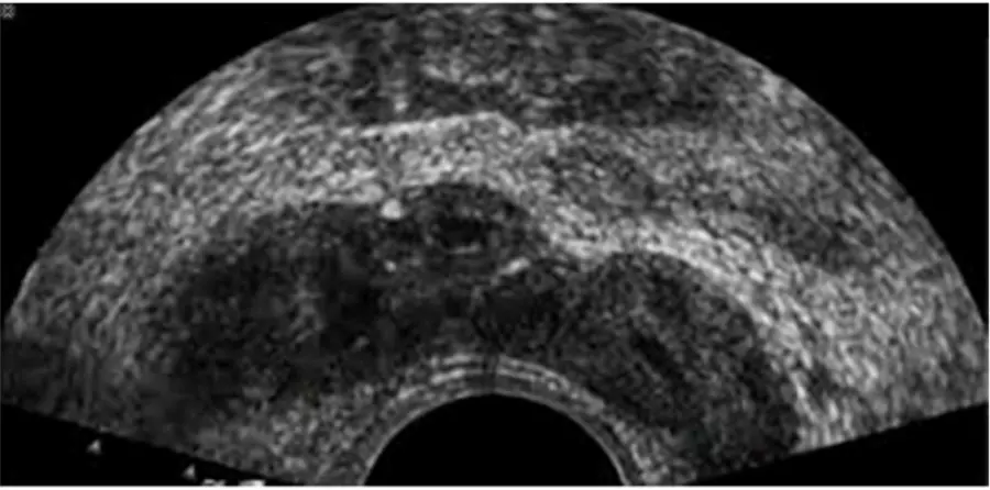
Which anatomical area of the male reproductive system is demonstrated in this endorectal image?

Correct Answer: C. Seminal vesicles
Explanation:

The ultrasound image shown is a transverse endorectal (transrectal) ultrasound, commonly used to evaluate the prostate and adjacent structures. The two hypoechoic (dark) oval-shaped structures seen superior and posterior to the prostate are characteristic of the seminal vesicles.

The seminal vesicles are paired, elongated glands located superior and posterior to the base of the prostate and are best visualized in transverse planes on endorectal imaging. They appear as hypoechoic or anechoic structures with internal septations, depending on the degree of fluid content.

In contrast:

The urethra appears as a central echogenic linear structure within the prostate.

The prostate base is more inferior in the scan and is visualized just above the urethra.

The ejaculatory ducts are usually not as prominently visualized and are located medial to the seminal vesicles, entering the prostate near the verumontanum.

This image most clearly demonstrates the bilateral seminal vesicles.


Rumack CM, Wilson SR, Charboneau JW, Levine D. Diagnostic Ultrasound, 5th ed. Elsevier; 2017.

ACR--AIUM--SRU Practice Parameter for the Performance of an Ultrasound Examination of the Prostate (2021).

Hagen-Ansert SL. Textbook of Diagnostic Sonography, 8th ed. Elsevier; 2017.

Question 15

Which sonographic finding indicates the need for immediate surgical intervention following testicular trauma?

Correct Answer: B. Discontinuity of the tunica albuginea
Explanation:

The tunica albuginea is a dense fibrous capsule surrounding the testis. Discontinuity of the tunica albuginea on ultrasound is diagnostic of testicular rupture --- a urologic emergency that requires immediate surgical repair to preserve testicular function and viability. Early surgical intervention within 72 hours has a high success rate for testicular salvage (up to 90%).

Intratesticular hematoma (A) may be managed conservatively if the tunica albuginea is intact.

Heterogeneity of the parenchyma (C) indicates injury but not necessarily rupture.

Increased vascularity (D) may be seen with inflammation or reperfusion but does not mandate surgery unless rupture is present.

Reference Extracts:

Dogra VS, Bhatt S. 'Acute painful scrotum: ultrasound evaluation.' Radiologic Clinics of North America. 2004; 42(2):349-363.

Middleton WD, Kurtz AB, Hertzberg BS. Ultrasound: The Requisites. 3rd ed. Elsevier, 2015.

---