
Limited-Time Offer: Enjoy 50% Savings! Ends in 00h 00m 00s Coupon code: 50OFF
Skip to content

Free ARDMS AE Adult Echocardiography Examination AE-Adult-Echocardiography Exam Questions

Page: 1 / 14 Total 139 questions

Want more questions? Get Premium Access.

Question 1

A "dropout" or loss of echoes from structures posterior to a calcified mitral annulus results in which artifact?

Correct Answer: C. Shadowing
Explanation:

Acoustic shadowing is the artifact caused by calcified structures like the mitral annulus, resulting in attenuation or loss of echoes from structures posterior to the calcification. The calcification absorbs or reflects the ultrasound waves, preventing them from reaching deeper structures and causing a 'dropout' or dark shadow behind the calcified area.

Reverberation involves repeated reflections creating multiple echoes. Side lobe artifacts arise from off-axis beams. Ring-down artifacts result from resonance in fluid or gas bubbles, not calcifications.

This artifact is explained in the 'Textbook of Clinical Echocardiography, 6e', Chapter on Ultrasound Artifacts20:75-80Textbook of Clinical Echocardiography.
Question 2

How are pseudoaneurysms best distinguished from true aneurysms?

Correct Answer: B. True aneurysms are lined with myocardium.
Explanation:

True ventricular aneurysms are lined by scarred myocardium and have a broad neck. Pseudoaneurysms occur after myocardial rupture contained by pericardium or scar tissue and lack myocardium in the wall. Pseudoaneurysms typically have a narrow neck and are more prone to rupture.

Pseudoaneurysms can occur at various locations, not exclusively the apex. Both true aneurysms and pseudoaneurysms may contain thrombus, so this is not a distinguishing feature.

This differentiation is important clinically and is detailed in adult echocardiography and surgical cardiology texts and ASE guidelines16:Textbook of Clinical Echocardiography, 6ep.400-40512:ASE Cardiac Masses and Aneurysms Guidelinesp.150-160.


Question 3

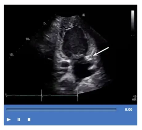
Which structure is the arrow pointing to in this video?

Correct Answer: C. Coronary sinus
Explanation:

The arrow points to the coronary sinus, which is a venous structure located posteriorly in the atrioventricular groove, emptying into the right atrium. It appears as a circular anechoic structure near the left atrium in echocardiographic images.

Left lower pulmonary vein enters the left atrium more superiorly. Descending aorta is posterior to the heart but not in this location. Left atrial appendage is an anterior finger-like projection of the left atrium, separate from the coronary sinus.

This anatomy is described in the 'Textbook of Clinical Echocardiography, 6e', Chapter on Cardiac Venous Anatomy20:140-145Textbook of Clinical Echocardiography.


Question 4

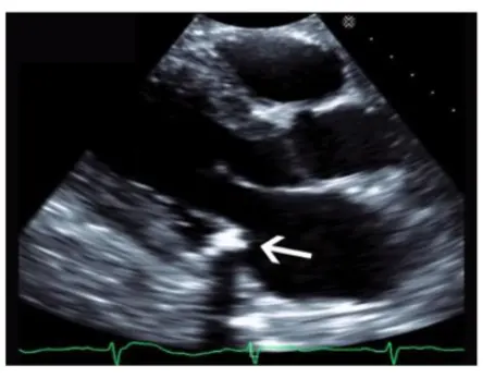
Which echogenic structure is indicated by the arrow on this image?

Correct Answer: D. Annular calcification
Explanation:

The image is a parasternal long-axis echocardiographic view focusing on the mitral valve annulus with a highly echogenic, dense, and well-defined structure located at the base of the posterior mitral leaflet. This appearance is characteristic of mitral annular calcification (MAC), a degenerative process resulting in calcium deposition along the mitral valve annulus.

Vegetations appear as irregular, mobile masses attached to valve leaflets and are less dense. Tumors and thrombi have different echogenicity and locations (tumors often in atria, thrombi in atrial appendages). MAC is usually more echogenic and localized to the annulus.

This description and differentiation are found in adult echocardiography textbooks and ASE guidelines on cardiac masses and valvular calcifications16:Textbook of Clinical Echocardiography, 6ep.460-46512:ASE Guidelines on Cardiac Massesp.150-160.


Question 5

Which maneuver aids in uncovering potential diastolic dysfunction while performing pulsed wave Doppler of the mitral valve?

Correct Answer: A. Valsalva
Explanation:

The Valsalva maneuver transiently reduces preload, which can unmask or exaggerate diastolic dysfunction during mitral inflow Doppler assessment. During Valsalva, changes in mitral E and A wave velocities can differentiate between normal and pseudonormal filling patterns by observing alterations in filling pressures.

Hand grip, leg air cycling, and squatting affect afterload and preload differently but are less specific for assessing diastolic dysfunction via mitral Doppler.

This technique is outlined in the 'Textbook of Clinical Echocardiography, 6e', Chapter on Diastolic Function Assessment and Maneuvers20:210-215Textbook of Clinical Echocardiography.


Question 6

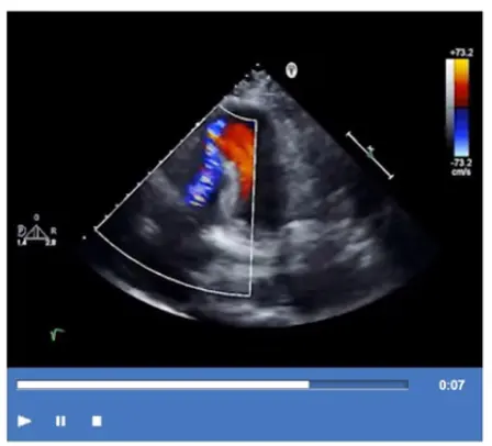
Which type of defect can be seen in this video clip?

Correct Answer: D. Ischemic ventricular septal defect
Explanation:

The echocardiographic video shows a defect in the ventricular septum with left-to-right shunting consistent with an ischemic ventricular septal defect (VSD), a mechanical complication of myocardial infarction. The defect allows blood flow between the left and right ventricles.

Coronary artery aneurysm appears as dilated coronary vessels, not a septal defect. Tricuspid regurgitation involves the right atrioventricular valve and is identified differently. Pseudoaneurysm of the apex is a contained myocardial rupture with narrow neck and does not involve septal communication.

This complication and its echocardiographic features are described in the 'Textbook of Clinical Echocardiography, 6e', Chapter on Post-Infarction Mechanical Complications20:430-435Textbook of Clinical Echocardiography.


Question 7

A continuous flow murmur is most likely due to which abnormality?

Correct Answer: B. Patent ductus arteriosus
Explanation:

A continuous murmur, heard throughout systole and diastole, is most characteristically caused by a patent ductus arteriosus (PDA). PDA represents persistent communication between the aorta and pulmonary artery, allowing continuous blood flow during both phases of the cardiac cycle.

Ventricular septal defect usually produces a holosystolic murmur. Concomitant aortic stenosis and mitral regurgitation cause separate murmurs but not continuous. Ebstein anomaly with atrial septal defect typically produces murmurs related to tricuspid regurgitation or ASD but not a continuous murmur.

This clinical correlation is detailed in the 'Textbook of Clinical Echocardiography, 6e', Chapter on Congenital Heart Disease and Murmur Etiologies20:420-425Textbook of Clinical Echocardiography.


Question 8

What is the range of the aortic valve area in normal adults?

Correct Answer: B. 3 - 4cm2
Explanation:

Comprehensive and Detailed Explanation From Exact Extract:

The normal aortic valve area (AVA) in adults typically ranges from 3 to 4 cm. This measurement is important for assessing aortic stenosis severity; values below this range suggest valve narrowing.

AVA values of 1-2 cm indicate mild to moderate stenosis, while less than 1 cm reflects severe stenosis. Larger areas like 5-6 or 7-8 cm are not physiologically typical.

This normal range is documented in the 'Textbook of Clinical Echocardiography, 6e', Chapter on Aortic Valve Anatomy and Function20:360-365Textbook of Clinical Echocardiography.


Question 9

Which statement is most accurate regarding cardiac contusion?

Correct Answer: A. It affects the right ventricle more commonly than the left.
Explanation:

Cardiac contusion is a myocardial injury resulting from blunt chest trauma, typically affecting the right ventricle more commonly than the left ventricle because of its anterior location and proximity to the chest wall. The injury can range from mild bruising to severe myocardial damage and dysfunction.

It does not result from myocardial infarction (which is ischemic injury), nor does it cause hypertrophy or hypercontractility. Instead, it may cause wall motion abnormalities, arrhythmias, or even rupture.

These features are detailed in echocardiography and trauma cardiology literature, including the 'Textbook of Clinical Echocardiography' and clinical guidelines on blunt cardiac injury16:Textbook of Clinical Echocardiography, 6ep.600-60512:ASE Trauma Cardiology Guidelinesp.500-505.


Question 10

Which method is appropriate for measuring the left atrial diameter in parasternal long axis?

Correct Answer: A. Inner edge to inner edge, perpendicular to the aortic root, at end-diastole
Explanation:

Comprehensive and Detailed Explanation From Exact Extract:

The recommended method to measure left atrial diameter in the parasternal long axis view is the inner edge to inner edge technique, perpendicular to the aortic root, measured at end-diastole. This approach provides the most reproducible and standardized measurement.

Measurement parallel to the aortic root or at end-systole is less accurate. Outer edge measurements overestimate size.

ASE chamber quantification guidelines specify this method for standardization and reproducibility in adult echocardiography practice12:ASE Chamber Quantification Guidelinesp.90-9516:Textbook of Clinical Echocardiography, 6ep.120-125.