
Limited-Time Offer: Enjoy 50% Savings! Ends in 00h 00m 00s Coupon code: 50OFF
Skip to content

Free ARDMS Sonography Principles and Instrumentation SPI Exam Questions

Page: 1 / 15 Total 215 questions

Want more questions? Get Premium Access.

Question 1

If the pulse repetition frequency is 3 kHz, what is the maximum Doppler shift that can be detected without aliasing?

Correct Answer: A. 1.5 kHz
Explanation:

The maximum Doppler shift that can be detected without aliasing is determined by the Nyquist limit, which is half of the pulse repetition frequency (PRF). If the PRF is 3 kHz, the Nyquist limit is 32=1.523kHz=1.5kHz. Therefore, the maximum Doppler shift that can be detected without aliasing is 1.5 kHz. Aliasing occurs when the Doppler shift exceeds this limit, causing an incorrect representation of the velocity. Reference: ARDMS Sonography Principles and Instrumentation, Chapter on Doppler Principles.
Question 2

Which feature is a characteristic of continuous wave Doppler?

Correct Answer: D. Dedicated transmit and receive crystals
Explanation:

Comprehensive and Detailed Explanation From Exact Extract:

Continuous wave (CW) Doppler utilizes two separate crystals: one constantly transmitting and one constantly receiving. This setup allows CW Doppler to measure very high velocities without aliasing, but at the cost of losing range specificity (no depth resolution).

According to sonography instrumentation reference:

''Continuous wave Doppler uses two separate elements for continuous transmission and reception of sound waves, allowing for detection of very high velocities but without depth specificity.''

Therefore, the correct answer is D: Dedicated transmit and receive crysta


Question 3

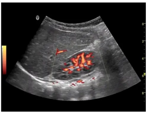
Which transducer was most likely used to create this image?

Correct Answer: A. Curvilinear
Explanation:

The image shown is typical of an abdominal ultrasound, which commonly utilizes a curvilinear transducer. Curvilinear transducers have a wider field of view at depth, making them ideal for imaging large structures within the abdomen. These transducers emit a curved beam, allowing for better penetration and a broader field of view, which is necessary for comprehensive abdominal examinations. The curvature of the image, the wide field of view, and the depth of penetration all suggest the use of a curvilinear transducer.


American Registry for Diagnostic Medical Sonography (ARDMS). Sonography Principles and Instrumentation (SPI) Examination Review Guide.

Question 4

Which characteristic of ultrasound transmission is directly proportional to an increase in frequency?

Correct Answer: C. Attenuation
Explanation:

Comprehensive and Detailed Explanation From Exact Extract:

As frequency increases, attenuation (energy loss due to absorption, scattering, and reflection) also increases. This limits penetration but improves resolution for superficial structures.

According to sonography instrumentation reference:

''Attenuation increases directly with frequency, limiting penetration but enhancing resolution in superficial imaging.''

Therefore, the correct answer is C: Attenuation.

---


Question 5

What parameter change reduces spectral Doppler aliasing?

Correct Answer: A. Increase Doppler angle
Explanation:

Comprehensive and Detailed Explanation From Exact Extract:

Spectral Doppler aliasing occurs when the Doppler frequency shift exceeds the Nyquist limit. The Doppler shift is proportional to the cosine of the Doppler angle. Increasing the Doppler angle (moving closer to 90 degrees) reduces the cosine value, thus reducing the Doppler shift frequency and helping to avoid aliasing.

According to sonography instrumentation reference:

''Increasing the Doppler angle reduces the Doppler shift frequency, thus decreasing the likelihood of aliasing in spectral Doppler.''

Therefore, the correct answer is A: Increase Doppler angle.

---


Question 6

Which method of sanitizing the transducer would damage piezoelectric crystals?

Correct Answer: D. Autoclave
Explanation:

Autoclaving involves high-pressure steam at high temperatures, which can damage the delicate piezoelectric crystals within the ultrasound transducer. These crystals are responsible for converting electrical energy into sound waves and vice versa. Exposure to the extreme conditions of an autoclave can cause thermal and mechanical damage to the crystals, rendering the transducer ineffective.


ARDMS Sonography Principles and Instrumentation guidelines

Zagzebski, J. A. (1996). Essentials of Ultrasound Physics.

Question 7

What is the primary factor that affects axial resolution?

Correct Answer: B. Spatial pulse length
Explanation:

Comprehensive and Detailed Explanation From Exact Extract:

Axial resolution refers to the ability to distinguish two structures that are located along the path of the ultrasound beam. It is directly related to the spatial pulse length (SPL), which is the product of the number of cycles per pulse and the wavelength. A shorter SPL leads to better axial resolution.

According to sonography instrumentation reference:

''Axial resolution improves as spatial pulse length decreases because shorter pulses can better differentiate structures lying close together along the beam axis.''

Therefore, the correct answer is B: Spatial pulse length.

---


Question 8

Which resolution is improved by focusing?

Correct Answer: A. Lateral
Explanation:

Focusing improves lateral resolution in ultrasound imaging. Lateral resolution refers to the system's ability to distinguish between two points that are side by side (perpendicular to the sound beam's path). By focusing the ultrasound beam, the width of the beam is narrowed at the focal point, enhancing the system's ability to resolve structures that are close together in the lateral plane. This results in clearer, more detailed images of the anatomical structures. Reference:

American Registry for Diagnostic Medical Sonography (ARDMS) Sonography Principles and Instrumentation study materials.

Diagnostic Ultrasound: Principles and Instruments by Kremkau, F. W. (latest edition).


Question 9

While imaging at a depth of 2 cm, which adjustment would improve the axial resolution?

Correct Answer: D. Increase frequency
Explanation:

Comprehensive and Detailed Explanation From Exact Extract:

Axial resolution improves with shorter spatial pulse length, which is directly related to higher frequency. At shallow depths (such as 2 cm), higher frequency can be used effectively since attenuation is minimal.

Principles and Instrumentation state:

'Axial resolution improves with increasing frequency due to shorter wavelength and pulse length.'

Turning off spatial compounding (A) affects speckle reduction.

Harmonics (B) help with resolution but primarily lateral contrast.

Gain (C) affects brightness, not resolution directly.

Therefore, the correct answer is D: Increase frequency.


Question 10

What reduces speckle and increases visualization of specular reflectors and attenuated structures?

Correct Answer: D. Spatial compounding
Explanation:

Comprehensive and Detailed Explanation From Exact Extract:

Speckle is a form of acoustic noise caused by interference of scattered echoes. Spatial compounding acquires multiple images from different angles and combines them to reduce speckle, enhance tissue texture, and improve visualization of structures that may otherwise be obscured by attenuation or artifact. The Principles and Instrumentation documentation states:

'Spatial compounding averages frames obtained at varying insonation angles. This reduces speckle artifact, smooths tissue texture, and improves visibility of specular reflectors and deeper structures.'

Therefore, the correct answer is D: Spatial compounding.

---


Question 11

Why is a higher frequency transducer a better choice for imaging superficial structures?

Correct Answer: D. Improved axial resolution
Explanation:

Comprehensive and Detailed Explanation From Exact Extract:

Higher frequency transducers provide better axial resolution due to their shorter wavelengths and spatial pulse lengths. This allows finer detail when imaging superficial structures where penetration is not a concern.

According to sonography instrumentation reference:

''Higher frequencies result in shorter pulse lengths, improving axial resolution and making them ideal for superficial imaging.''

Therefore, the correct answer is D: Improved axial resolution.

---


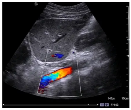
Question 12

What does this image demonstrate?

Correct Answer: C. Color aliasing
Explanation:

Color aliasing in Doppler ultrasound occurs when the velocity of blood flow exceeds the Nyquist limit, causing the color display to wrap around and display high velocities incorrectly as the opposite direction. This phenomenon is characterized by a mix of colors that indicate flow in both directions at the same location. In the provided image, there is a clear presence of color aliasing, as evidenced by the abrupt color change across the vessel, which is not consistent with normal flow patterns. Reference:

American Registry for Diagnostic Medical Sonography (ARDMS) Sonography Principles and Instrumentation guidelines.


Question 13

Which change would lead to a reduction in frame rate?

Correct Answer: A. Increasing the number of focal zones
Explanation:

Comprehensive and Detailed Explanation From Exact Extract:

Each additional focal zone requires extra transmissions per scan line, which slows down frame acquisition, thereby reducing the frame rate.

Principles and Instrumentation state:

'Increasing the number of focal zones lowers frame rate because multiple pulses are needed per scan line for each focal depth.'

Increasing PRF (B) allows faster data acquisition.

Decreasing depth (C) shortens round-trip time, increasing frame rate.

Decreasing sector width (D) reduces scan lines, increasing frame rate.


Question 14

Which type of resolution will be improved by decreasing the depth of field?

Correct Answer: A. Lateral
Explanation:

Lateral resolution refers to the ability to distinguish two structures that are side by side. It is dependent on the width of the ultrasound beam. By decreasing the depth of field, the beam width is reduced at any given point along the depth, which improves the lateral resolution. This is because a narrower beam can better distinguish between objects that are close together laterally.


ARDMS Sonography Principles and Instrumentation guidelines

Kremkau, F. W. (2015). Diagnostic Ultrasound: Principles and Instruments.

Question 15

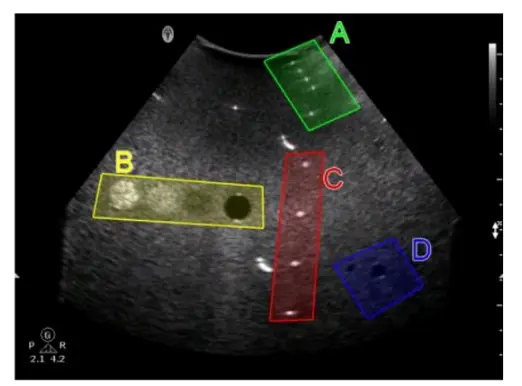
Which target group in this image of a tissue-mimicking phantom is used for gray-scale evaluation?

Correct Answer: C. Option C
Explanation:

Gray-scale evaluation in a tissue-mimicking phantom involves assessing the uniformity and accuracy of the gray-scale representation of the tissues.

Option C typically contains structures designed to test the machine's ability to accurately depict varying levels of echogenicity, which is essential for proper gray-scale evaluation.

This area will have a range of echo intensities that help in determining the contrast resolution and the ability of the system to distinguish between different tissue types based on their gray-scale values. Reference:

ARDMS Sonography Principles and Instrumentation guidelines on tissue-mimicking phantoms and image quality evaluation.